

19 Oct Anatomage Table Arrives at the University of Dayton


Students at the University of Dayton will soon be able to delve into the human body and uncover the workings of blood vessels, nerves and more thanks to a new 3D virtual anatomy visualization and virtual dissection system. The Anatomage Table is an important addition to the department of health and sport science during COVID-19; the department plans to use the technology to support students who are learning remotely to continue a high-quality, cadaver-based laboratory educational experience.

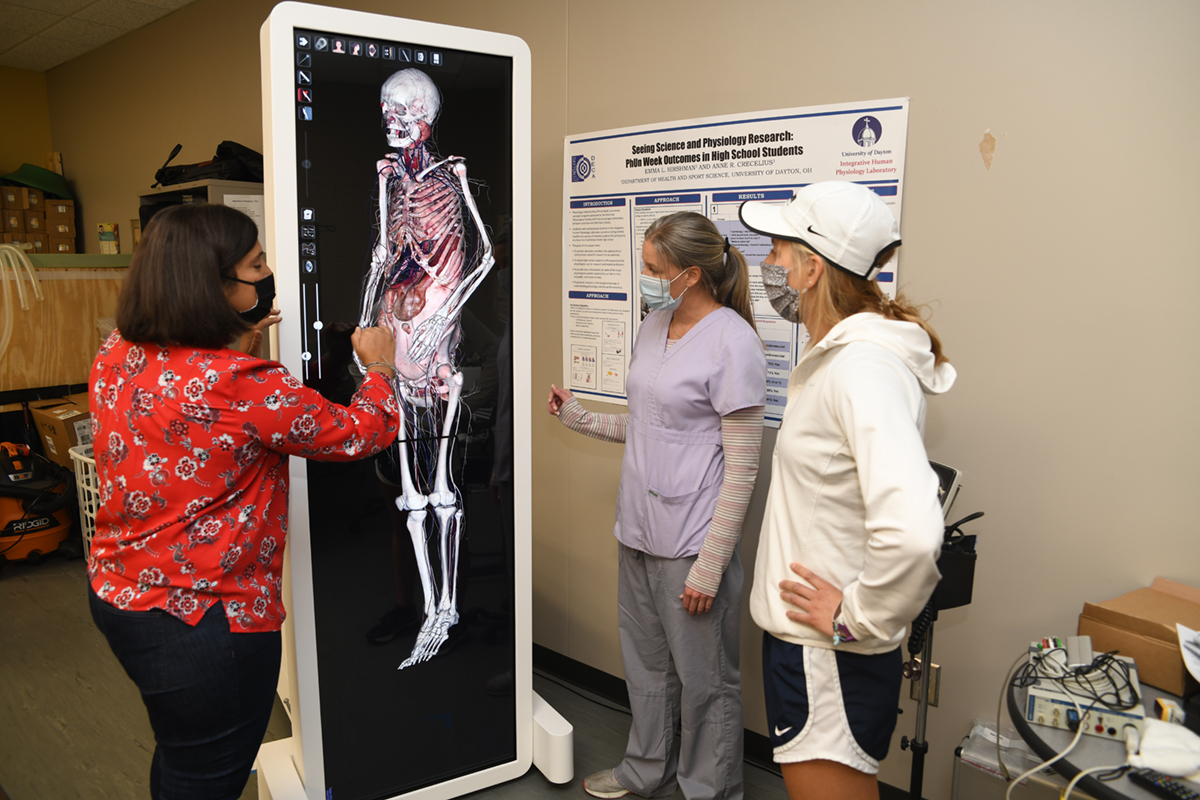
(from left) Anne Crecelius, Kim Ritterhoff, and Student Kelly Wilker use the anatomy table. The program on the table is for gross anatomy. The table will help students see organs, various systems, like circulatory system, and case studies.
“A lot of graduate clinical programs are adopting similar technology, so our undergraduate students will have the experience of using this technology before starting advanced coursework,” said Kim Ritterhoff, human anatomy and human dissection lecturer in the School of Education and Health Sciences. “It should give them more experience and allow them to be ready for their next level.”
The Anatomage Table, the size of a human body, allows students to examine, study and dissect 3D structures as if they were in a lab with a real cadaver. Coursework will allow students to understand the structures and functions of living bodies. The table also includes CT and MRI software. It also will offer a chance for students to learn about the anatomical changes that occur through aging and various diseases and conditions as well as view the structural aspects of surgical repairs and interventions. This allows them to become more familiar with the variety of clinical situations they may encounter in their future professions.
It was made possible thanks to a gift from David and Norma McCarthy, 1971 graduates of the University of Dayton.